- admin
- 0 Comments
For much vascular pathology, ultrasound has established itself as a method of diagnosis allowing the physician to assess the blood flow and blood vessels without surgery as it is safe and precise. There is a need for a thorough evaluation of vascular diseases such as deep vein thrombosis DVT, peripheral artery disease PAD, varicose veins, and aneurysms among others when formulating treatment regiments. Thanks to innovative technological advancement in ultrasound, healthcare professionals can directly see the blood flow and blood vessel operation and take any necessary actions as barriers, thrombi or even frail vessel walls can be observed.
In this article we will look at the significant role of ultrasound in the assessment of vascular pathology, its advantages and how it assists in the treatment of such conditions.
What is Vascular Ultrasound?
Vascular ultrasound, or Doppler ultrasound in some contexts, is a type of sonography used for examinations of extremity blood vessels and the abdominal aorta. This principle enables measurement of movement of blood by evaluating how the sound waves are presented through the moving, and reflected by, today’s blood cells orientated in the fetus. It is a short and painless activity which is done in physician office or hospital or imaging centers.
Ultrasound is useful to evaluate several vascular problems such as:
- Occlusion or narrowing of the arteries (stenosis)
- Thrombus in venous blood such as in cases of Deep vein thrombosis (DVT)
- Bulging and weakening of a blood vessel: Aneurisms
- Spider veins or valvular insufficiency in veins
- Ischemia of the lower extremities caused by peripheral artery disease (PAD)
What purpose does vascular ultrasound serve?
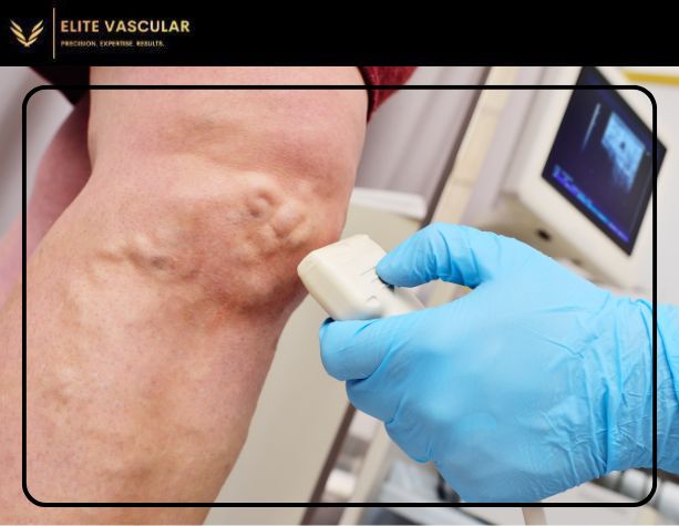
Vascular ultrasound is the use of sound waves that pass through a device known as the transducer, which is a hand held device placed on the skin. The transducer generates sound waves that are directed to the human body and the tissues and blood cells present within the body. These sound waves which are reflected back from the tissues and blood cells are picked up by the monitor cum machine, which then displays them as pictures or graphs representing the movement of blood and the shape of the vessels.
Banking on different colors and shades is one of the most fundamental in vascular ultrasound; this effect contributes to the velocity of blood flow moving towards the ear and away from the ear even without the use of any monitor. This is particularly important in the investigation of vascular parasites such as the blockage in the arteries that hinders the supply of blood or the case where an oversupply of blood to the heart is experienced where an inefficient venous system does not allow blood to be efficiently transported back to the heart.
Types of Vascular Ultrasound
Vascular ultrasound does not only that but has additional diagnostic functionalities and can be divided into several classes, in which the devices are applied:
- Duplex ultrasound: the module combines standard ultrasound and Doppler module in order to obtain not images only or direction and speed of blood flow.
- Color Doppler: In color Doppler imaging, color is added in the Doppler ultrasound to measure the amount and direction of blood flow in the body.
- Continuous Wave Doppler: It measures blood flow velocity through the use of continuous sound waves, Useful in identifying fast blood flow in small arteries with narrow openings.
Diagnosing Routine Vascular Disorders by the help of Ultrasound Images
Deep Vein Thrombosis (DVT)
With the utilization of ultrasound, it is easy to confirm the diagnosis of deep vein thrombosis (DVT). This is a condition where the blood is trapped in the deep veins of the leg, in particular the muscles behind the calf and knee, due to an occlusive clot. If not diagnosed and treated swiftly, DVT can cause severe and fatal risks such as pulmonary embolism, a clot that dislodges and induces blockage in the lungs. Ultrasound imaging helps after surgery to evaluate for the presence of clots or to rule out the size or place of the clot and thus determine optimal treatment.
Peripheral Artery Disease (PAD)
The disease is most prevalent in older patients and is associated with the narrowing or blocking of arteries in the legs due to atherosclerosis limiting the blood supply to the limbs. Doppler ultrasound assists in locating the impaired blood flow areas and in evaluating the degree of occlusions. White measuring velocity, eg. quantifying blood flow, doctors can tell if the patient is indicated for such actions as angioplasty, stenting or other procedures meant for increasing blood inflow into the organs.
Varicose Veins
Dilated or swollen veins are known as varicose veins and are caused by malfunctions of the valves in the veins that lead to backward blood flow and pooling of blood. Doctor’s utilization of ultrasound helps determine the degree of valve incompetence in conjunction to the Doppler evaluation of blood flow in varicose veins, and plan the possible procedures and scripts such as sclerotherapy or vein ablation.
Aneurysms
In vascular diseases, this area can develop and bulge outwards. It may rupture and leads to severe complication. Ultrasound is used to locate such inflammation in large arteries like the aorta so that their growth can be followed and timely treatment can be implemented if needed.
Carotid Artery Disease
The carotid arteries carry blood to the brain and may get narrowed by plaque deposition which may put them at an increased risk of getting a stroke. Sonographic evaluations of the carotid arteries have been shown to be useful in detecting causes of cervical vascular disease to determine if operations such as carotid endarterectomy or angioplasty are required.
Benefits of Vascular Ultrasound
Moreover, a vascular ultrasound has several benefits in diagnosis of vascular pathology;
Non-Invasive and Painless
In contrast to other diagnostic modalities, vascular evaluation by ultrasound does not involve the use of contrast media, ionizing radiation or surgical interventions, and therefore can be considered as a safe procedure free of pain.
Live imaging
Ultrasound has the advantages of being a bedside imaging tool since the results are instant. Doctors are able to know the status of blood vessels and blood flow. This makes it very useful in the management of sudden vascular challenges such as DVT.
No Radiation Exposure
This makes ultrasound a better option as it does not use any ionizing radiation which may be dangerous to patients especially those undergoing clear surveillance like patients with chronic vascular conditions.
Portable and Accessible
Today, vascular ultrasound machines are quite small and can be used in all health institutions by many types of users as opposed to being used in specialized vascular sonographers’ areas only. This ensures that patients do not have to wait long for diagnosis and treatment most especially during emergencies.
Cost Efficient
Ultrasound has proven to be an economical form of diagnostic approach in the assessment of vascular health rather than carrying out the more invasive procedures which are more expensive.
When Is It Appropriate to Get Vascular Ultrasound done?
A vascular ultrasound may be recommended by your medical practitioner if you are at risk or presently have any symptoms of vascular disease such as: A vascular ultrasound may be recommended by your medical practitioner if you have risk factors or symptoms of vascular disease.
- Pain or cramping of the legs when engaging in physical activities (indicates PAD)
- Leg swelling or pain (Such as possibly DVT)
- Varicose veins or persistent leg swelling that may be not well understood
- High blood pressure or high blood cholesterol
- A self history or a family history of aneurysms or other related vascular conditions
Those who suffer from morbidity conditions such as PAD and degeneration of the leg veins which requires regular assessment with repetitive vascular ultrasound are the ones who do this so that they monitor how far their delaying strategies have worked and thus alter/maintain the course of treatment accordingly.
Conclusion: The Key Importance of Ultrasound in Vascular Health
Diagnosis and treatment of numerous vascular diseases, vascular ultrasound is an integral part in their management. Doctors use ultrasound technology to view and diagnose various blood vessel abnormalities ranging from blood clots to blockages in the arteries from cranial Aneurysms to varicose veins. The simplicity and cost efficiency does not compromise the problem solving accuracy making ultrasound an essential appliance in vascular disease diagnosis. In case you develop symptoms such as dull ache in the legs, discolored bulging veins in the feet, or discomfort when moving about consider reporting to your physician to check if a vascular ultrasound is an appropriate diagnostic measure for you.
